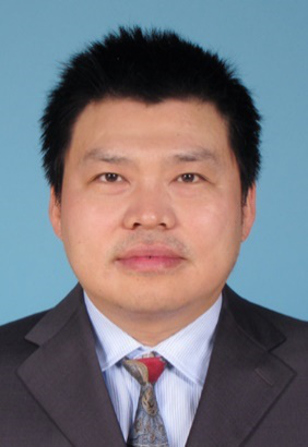
【报告题目】:活体高时空分辨率成像(High spatiotemporal resolution fluorescence imaging of biological samples in vivo)
【报告人】 北京大学特聘教授 陈良怡
【报告时间】 10月28日(周三)上午 9:30
【报告地点】 生物医学工程高精尖创新中心(窦店园区)2号楼2层西侧阶梯会议室
【邀请人】 生物医学工程高精尖创新中心医用光子学研究所

【陈良怡教授简介】
陈良怡 北京大学分子医学研究所博雅特聘教授、博士生导师,北京大学麦戈文脑研究所、北京智源人工智能研究院、北京市协同创新研究院兼职研究员,获国家自然科学基金委杰出青年基金、优秀青年基金资助。主要研究方向是糖尿病发病过程中胰岛素分泌异常机制,发明了一系列的高时空分辨率生物医学成像的可视化手段,包括:1) 高时空分辨微型化双光子显微镜,开创高分辨率在体成像领域。获2017年中国科学十大进展等奖项,入选Nature Methods 2018年年度方法;2) 高分辨率双光子光片显微镜,斑马鱼在体单个胰岛细胞成像并揭示其功能成熟机制;3) 超灵敏海森结构光显微镜,首次解析线粒体内嵴等结构动态变化,获2018年中国光学十大进展;4) 提出并发明活细胞双模态超分辨率显微镜,观察细胞器互作全景并发现新型细胞器。
【报告摘要】
Here we will present three pieces of high-resolution fluorescence microscopy methods we invented for live sample imaging. The first one is for in vivo imaging, which is a fast, high-resolution, miniaturized two-photon microscope (FHIRM-TPM). With a headpiece weighing 2.15 g and a new type of hollow-core photonic crystal fiber to deliver 920-nm femtosecond laser pulses, the FHIRM-TPM is capable of imaging commonly used biosensors at high spatiotemporal resolution (0.64 μm laterally and 3.35 μm axially, 40 Hz at 256 × 256 pixels). It compares favorably with benchtop two-photon microscopy and miniature wide-field fluorescence microscopy in the structural and functional imaging of Thy1-GFP- or GCaMP6f-labeled neurons. Further, we demonstrate its unique application and robustness with hour-long recording of neuronal activities down to the level of spines in mice engaging in social interaction. The second method is for live cell long-term super-resolution (SR) imaging. We have developed a deconvolution algorithm for structured illumination microscopy based on Hessian matrixes (Hessian-SIM). It uses the continuity of biological structures in multiple dimensions as a priori knowledge to guide image reconstruction and attains artifact-minimized SR images with less than 10% of the photon dose used by conventional SIM while substantially outperforming current algorithms at low signal intensities. Hessian-SIM enables rapid imaging of moving vesicles or loops in the endoplasmic reticulum without motion artifacts and with a spatiotemporal resolution of 88 nm and 188 Hz. Its high sensitivity allows the use of sub-millisecond excitation pulses followed by dark recovery times to reduce photobleaching of fluorescent proteins, enabling hour-long time-lapse SR imaging in live cells. The third technology is a dual-mode SR microscopy for highlighting molecules as well as a holistic view of related interacting organelles in live cells. It is a combination of two-dimensional Hessian-SIM with label-free three-dimensional optical diffraction tomography (ODT), term SR fluorescence-assisted diffraction computational tomography (SR-FACT). The ODT module is capable of resolving mitochondria, lipid droplets, the nuclear membrane, chromosomes, the tubular endoplasmic reticulum and lysosomes. Using dual-mode correlated live cell imaging for prolonged period of time, we observe a novel subcellular structure named dark-vacuole bodies, the majority of which originates from densely populated perinuclear regions and intensively interacts with organelles including mitochondria and the nuclear membrane, before ultimately collapsing into the plasma membrane. These works demonstrate the unique capabilities of SR-FACT, which suggest its wide applicability in cell biology in general.